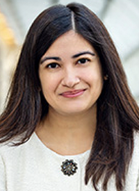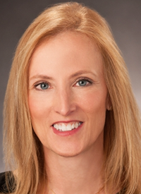

Reshma Jagsi, MD, DPhil, eagerly anticipated Friday afternoon at the 40th Annual Miami Breast Cancer Conference®, an event hosted by Physicians’ Education Resource® (PER®). That time was dedicated to radiation oncology—her specialty; it is both the depth and the breadth of the program that she finds exciting.
“I [couldn’t] wait for Friday afternoon because I’m a radiation oncologist,” said Jagsi, who is acting professor and chair of the Department of Radiation Oncology at Emory University School of Medicine in Atlanta, Georgia, in an interview with OncLive®. “But the whole program is just packed with interesting stuff. Breast cancer management is not just multidisciplinary but interdisciplinary. That is what I love about the Miami Breast Cancer [Conference]. There [were] sessions throughout the program that I [was] raptly listening to.”


Similarly, Kelly K. Hunt, MD, emphasized her takeaways from the meeting each year. Hunt gave the presentation in Saturday’s surgical track called “Breast-Implant Associated Anaplastic Large Cell Lymphoma: What Have We Learned?”
“One of the things I always say is, ‘After the conference, what are you going to do on Monday morning? How are you going to alter or change your practice?’” said Hunt, who is the Olla Stribling Distinguished Chair for Cancer Research at The University of Texas MD Anderson Cancer Center in Houston. “[The organizers] try to give pearls of wisdom throughout the conference about how these new treatments are going to [affect] patients, how they get incorporated, [and] how to educate our patients on different therapies. Overall, it’s a fun conference to participate in but also to be in the audience and hear some of the updates.”
Elizabeth Anne Morris, MD, professor and chair of the Department of Radiology at UC Davis Health in Sacramento, California, echoed those comments. Her presentation, “Practice Patterns in the Use of MRI in Primary Operable Breast Cancer,” was scheduled for Friday afternoon’s surgery track. The cross-training that takes place throughout the conference makes perfect sense to her.
“Radiology and surgery work hand in hand. In taking care of patients [with] breast cancer, we rely on each other and work in tandem,” she said. “It’s a very close collaboration. That’s one area that I truly enjoy in the clinic: working with my surgical colleagues to get the best outcomes for our patients.”
MRI augments the radiologist’s training and experience with deep learning techniques to evaluate whether a patient has experienced complete pathologic response. She said the combination makes imaging and testing more sensitive and specific for each patient and provides better information for the surgeon and radiation oncologist. The growth of artificial intelligence (AI) allows physicians to see tumors in unprecedented ways.
AI, she said, is a broad term that includes techniques such as radiomics, machine learning, and deep learning, which is a subset of machine learning. The MRI produces not only pictures alone but pictures that comprise data; AI can analyze data in those images that are invisible to the human eye. The combination of AI and a skilled radiologist working together can better assess those data and determine whether a patient has responded to treatment. “It’s through not only using tools that assess for the spatial relationships between pixels but also tools that use large data sets where you train these convolutional neural networks to recognize patterns to assess whether or not a response has happened,” Morris said.
In data published in 2018, investigators assessed the effect of whole-body MRI (WB-MRI) added to CT of chest-abdomen-pelvis (CT-CAP) and 18F-fluorodeoxyglucose PET (positron emission tomography)/CT on systemic treatment decisions for patients with advanced breast cancer. Among 910 WB-MRI examinations, 58 had a paired control examination (16 CT-CAP and 42 PET/CT).1 WB-MRI detected additional sites of disease in 23 paired examinations; additional sites of disease were not detected in the control examination. In 17 paired examinations, WB-MRI detected progressive disease that was not detected in the control examination.
Investigators changed treatment in 14 of 28 pairs of examinations after WB-MRI identified progressive disease. The control examination only detected stable disease in those patients.1
In another study, investigators at Duke University in Durham, North Carolina, conducted a retrospective analysis of dynamic contrast-enhanced fat-saturated T1-weighted MRI sequences from 131 patients with a diagnosis of ductal carcinoma in situ (DCIS) confirmed by core needle biopsy.2 They used 2 different deep learning approaches to predict whether there was an occult invasive component in the analyzed tumors identified at surgical excision.
In the first approach, they used the pretrained GoogLeNet transfer learning strategy. In the second, they used a pretrained network to extract deep features and a support vector machine (SVM) that uses these features to predict the upstaging of DCIS. They validated their findings using nested 10-fold cross-validation and the area under the receiver operating characteristic curve (AUC) to estimate the performance of the predictive models.
The GoogLeNet model pretrained on ImageNet as the feature extractor and a polynomial kernel SVM used as the classifier produced the best classification performance (AUC, 0.70; 95{61098da95f7e9566452289a1802d8d1a52c0e4ce3811e4bc55deae57fae5622a} CI, 0.58-0.79). The highest AUC obtained with the transfer learning–based approach was 0.68 (95{61098da95f7e9566452289a1802d8d1a52c0e4ce3811e4bc55deae57fae5622a} CI, 0.57-0.77).2
Investigators concluded that deep learning methods applied to preoperative breast MRIs in women with newly diagnosed DCIS could predict occult invasive disease. Results were “relatively good” using the off-the-shelf GoogLeNet approach, which “shows promise” for future testing. On the other hand, they found that the transfer learning–based approach resulted in overfitting and poor classification performance.
Morris added that MRI can be used for screening and in the diagnostic setting to differentiate benign vs malignant tumors. The potential of MRI in the neoadjuvant setting intrigues her because more women with breast cancer are undergoing neoadjuvant treatment and many of them have exceptional responses.
“Now that we have more targeted therapies, we’re able to see these complete responses on imaging,” Morris said. “That really opens the door to consider possibly one day [managing] breast cancer with therapy and assessing with an MRI examination. These exceptional responders may not need surgery. That would be converting something that has always been a surgical disease to something that is no longer a surgical disease, and that is exciting to me.”
Less Radiation Treatment Is the Goal
Although MRI may someday increase the pool of patients with breast cancer who can avoid surgery, Jagsi is working to reduce—and in some cases, eliminate—the need for radiation therapy. Aside from the modality’s effectiveness, it also causes toxicity, she said. Over the past 2 decades, the trend in clinical practice has moved toward using less radiation whenever possible. “It has costs, so when we balance the benefits of radiation against those costs, we seek ways to make the radiation treatment less burdensome or less toxic or we seek to identify patients in whom other treatments have made the condition so well controlled that we might safely omit radiation treatment altogether,” she said.
Jagsi discussed ways to deescalate or omit radiation therapy in the plenary session held on Friday morning. Improved screening techniques can identify breast cancer earlier than ever before, she said. Surgeons are performing more precise resections, pathologists are improving their process of specimens, and advances in systemic therapy allow physicians to tailor their treatments more precisely to an individual patient. All these developments create opportunities to potentially deescalate radiation therapy, she said.
“When we say deescalation, there is a variety of ways we can deescalate treatment. We can omit it altogether in certain patients or we can shorten the course of treatment or deintensify the dose,” Jagsi said. “There have been a number of efforts in place to look at whether we really need to treat most patients.”
In July 2022, investigators analyzed records from 631 patients 70 years or older with hormone-sensitive, pT1N0M0 invasive breast cancer treated with endocrine therapy. Forty-seven patients (7.4{61098da95f7e9566452289a1802d8d1a52c0e4ce3811e4bc55deae57fae5622a}) met the criteria for radiation omission and were treated by 14 surgeons at 8 institutions.3
Thirty-four of the 47 patients (72.3{61098da95f7e9566452289a1802d8d1a52c0e4ce3811e4bc55deae57fae5622a}) eligible for omission ultimately received adjuvant radiation. Those who received radiation were significantly younger than those who did not (P = .03). Investigators found no difference in radiation use based on size (P = .40), histology (P = .50), grade (P = .70), race (P = .10), ethnicity (P = .60), institution (P = .10), gender of the surgeon (P = .70), or surgeon (P = .10).
Jagsi warned that treatment decisions are complex, with patients who are eligible for omission or even deescalation choosing to undergo radiation therapy for any number of reasons. Radiation can reduce the risk for local recurrence, some patients may be unable or unwilling to undergo endocrine therapy, and improvements in delivery have significantly reduced toxicity. “What’s really important is to make sure that all [patients] who are candidates for omission of radiation [and] who have relatively low benefits as well as relatively low risks from treatment are able to make decisions for themselves that are concordant with their own values and their own preferences,” she said.
Jagsi recognized the importance of interdisciplinary collaboration, but she was still most excited about the presentations on radiology and radiation oncology scheduled for the conference. She was looking forward to a presentation by Fleure Gallant, MD, a radiation oncologist at Maimonides Cancer Center in Brooklyn, New York, that will explore whether bolus (where physicians apply a layer tissue on top of the chest wall following mastectomy) is grounded in evidence and appropriate in the modern era of breast cancer management.
Timothy J. Whelan, BSc, BM, BC, the Canada Research Chair in Breast Cancer Research at McMaster University in Hamilton, Ontario, Canada, spoke about nodal irradiation on Friday afternoon. Jagsi said this is an area where physicians are interested in identifying patients most likely to benefit and using a more sophisticated understanding of biology to make those decisions. “The radiation treatment lineup is a star-studded cast with interesting presentations,” she said.
Embrace Debate
Hunt is slated to give presentations on breast implant–associated anaplastic large cell lymphoma and axillary reverse mapping (ARM), a technique developed to prevent lymphedema. Data from a systemic literature review published in January 2023 showed that pooled lymphedema incidence in the ARM group was 4.8{61098da95f7e9566452289a1802d8d1a52c0e4ce3811e4bc55deae57fae5622a} compared with 18.8{61098da95f7e9566452289a1802d8d1a52c0e4ce3811e4bc55deae57fae5622a} in patients who received conventional axillary dissection (P < .0001).4
Hunt said she was looking forward to a presentation by Laura J. Esserman, MD, MBA, director of the Carol Franc Buck Breast Care Center at the University of California, San Francisco School of Medicine and the 2018 Giants of Cancer Care® award winner in cancer diagnostics. Esserman will provide an update on data from the I-SPY trials, results Hunt called “incredibly valuable.”
Jagsi said she was anticipating a Medical Crossfire® session between Atif Khan, MD, MS, enterprise-wide director of breast radiotherapy services at Memorial Sloan Kettering Cancer Center in New York City, New York, and Jean L. Wright, MD, associate professor of radiation oncology and molecular radiation sciences at Johns Hopkins University in Baltimore, Maryland. Like Jagsi, the Medical Crossfire debates are a highlight of the conference. Jagsi debated Tari A. King, MD, FACS, chief of the Division of Breast Surgery at Dana-Farber Cancer Institute in Boston, Massachusetts, on targeted axillary dissection.
“We all read data or studies and interpret the data with our own viewpoint,” Jagsi said. “It’s always good to hear what others have to say about how the studies were designed and what weaknesses there might be in the data analysis and interpretation,” Hunt said. “When you hear these Medical Crossfires, I think people are always trying to poke holes in in the data and show you where there are weaknesses or strengths that someone else might not have realized.”
References
- Zugni F, Ruju F, Pricolo P, et al. The added value of whole-body magnetic resonance imaging in the management of patients with advanced breast cancer. PLoS One. 2018;13(10):e0205251. doi:10.1371/journal.pone.0205251
- Zhu Z, Harowicz M, Zhang J, et al. Deep learning analysis of breast MRIs for prediction of occult invasive disease in ductal carcinoma in situ. Comput Biol Med. 2019;115:103498. doi:10.1016/j.compbiomed.2019.103498
- Hong MJ, Lum SS, Dupont E, et al; SHAVE2 Investigators. Omission of radiation in conservative treatment for breast cancer: opportunity for de-escalation of care. J Surg Res. 2022;279:393-397. doi:10.1016/j.jss.2022.06.036
- Co M, Lam L, Suen D, Kwong A. Axillary reverse mapping in the prevention of lymphoedema: a systematic review and pooled analysis. Clin Breast Cancer. 2023;23(1):e14-e19. doi:10.1016/j.clbc.2022.10.008


More Stories
‘I hope it gives young people some ideas!’: David Hockney’s immersive art show – photo essay | David Hockney
The Overall Winner of The Architecture Drawing Prize
10 Online Drawing Games To Play With Your Friends